Services
Kidneys

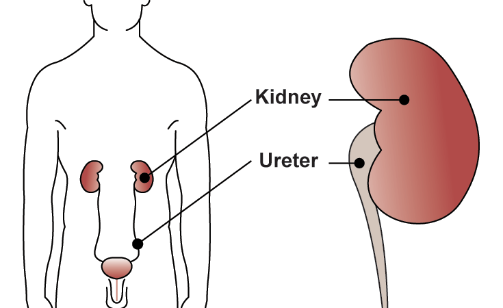
The kidneys are two bean-shaped body organs found behind the 13th rib. Their primary role is to help maintain the body in a state of balance by controlling the make-up and volume of blood. They remove wastes from the blood in the form of urine, and conserve water.
To maintain the necessary fluid balance, the kidneys filter 160 liters (roughly 42 gallons) of blood a day, but they excrete in the urine only about 1percent of the filtrate's salt and water content. This is accomplished by a very complex set of structures in the kidney called nephrons that delicately balance filtration, re-absorption, and secretion of various constituents of body fluid.
Water makes up approximately 95 percent of the total volume of urine, with the remaining 5 percent consisting of dissolved solutes, or wastes (i.e. urea, creatinine, and uric acid.) Urine is steadily excreted from the kidneys then passes down two ureters to the bladder by means of muscle contractions and the force of gravity. Once in the bladder, the urine is temporarily stored until it is voided from the body through the urethra.
The Lower Urinary Tract
The lower part of the urinary tract consists of a hollow muscular storage organ called the bladder, that is found in the pelvis behind the pelvic bone (pubic symphysis) and a drainage tube, called the urethra, that exits to the outside of the body. The urinary bladder, ureters, and urethra are all muscular structures lined with a membrane coated with mucus that is impermeable to the normal soluble substances of the urine.The urinary bladder is an elastic organ that changes shape according to the amount of urine it contains. It resembles a deflated balloon when empty but becomes somewhat pear-shaped and rises into the abdominal cavity when the amount of urine increases.
The bladder wall has three main layers of muscle: the mucosa, submucosa, and detrusor muscle. The mucosa is the innermost layer and is composed of transitional cell epithelium. The submucosa lies immediately beneath the mucosa and its basement membrane. It is composed of blood vessels that supply the mucosa with nutrients and the lymph nodes that aid in the removal of waste products. The detrusor is a thick layer of smooth muscle that expands to store urine and contracts to expel urine. The urethra is a small tube that leads from the floor or neck of the urinary bladder to the outside of the body. In women, the urethra is approximately 1.5 inches long and is found in the front wall of the vagina. The urethral orifice or meatus is the outside opening of the urethra and is located between the clitoris and the vaginal opening.
In men, the urethra is approximately 8 inches long. When it leaves the bladder, it passes downward through the prostate gland, the pelvic muscle and finally through the length of the penis until it ends at the urethral orifice or opening at the tip of the glans penis.
Storage and emptying of the bladder are regulated by the internal and external urethral sphincters. Sphincters are made up of a ring-like band of muscle fibers close off a natural opening in the body. Sphincters are normally in a closed position and need stimulation to open. Continence depends on two factors: normal lower urinary tract support and normal sphincteric function.
Lying below the internal sphincter is the external sphincter, which is made up of smooth muscle mixed with striated, or striped, muscle of the pelvic floor or pelvic diaphragm. Unlike the smooth muscles that an individual cannot consciously control, the striated muscles of the external sphincter allow for voluntary interruption of abdominal pressure to prevent urine leakage, such as occurs in coughing or sneezing.
These three sets of muscles must work in close unison to control the various stages of urinary bladder filling and emptying. During the filling stage, only minimal activity is needed to produce closure of the external urethral sphincter. At a certain point during bladder filling, the internal pressure within the bladder becomes strong enough to activate stretch receptors in the bladder wall. When these stretch receptors send a message to the nervous system, small contractile waves occur in the detrusor muscle and the internal urethral sphincter automatically relaxes and becomes funnel shaped. The external sphincter must now be consciously tightened, and the urge to urinate becomes very apparent. To urinate, a person must relax the external sphincter.
The advantage of this system is that, during the early stages of bladder filling, a person remains unaware of the slowly accumulating urine and is not required to keep the external sphincter tightly closed. This only becomes necessary when enough urine collects to relax the internal sphincter.
Pelvic Muscle Support
The mechanism responsible for supporting the bladder neck and urethra involves the interconnections of three structures: (1) the arcus tendinous fasciae pelvis, (2) the levator ani muscles, and (3) the endopelvic fascia around the urethra and vagina. The arcus tendinous fasciae pelvis is a fibrous band that is attached in the front to the pubic bone and in the back to the ischial spine. The endopelvic fascia is a group of connective tissue that connects the urethra and anterior vaginal wall. The levator ani is connective tissue that provides support to the urethra and vagina. It is made up of Type I, slow twitch muscle fibers and Type II, fast twitch muscle fibers. At least 80 percent of the levator ani muscle are Type 1 muscle fibers. These fibers produce less force on muscle contraction and assist in improving muscle endurance by generating a slower more sustained but less intense muscle contraction. Type I muscle fibers are also fatigue resistant.The second group is Type II or fast twitch fibers, which aid in quick and forceful contractions. These fibers are used during sudden increases in intra-abdominal pressure by contributing to closing of the urethra. Exercising Type II muscle fibers will increase muscle strength. Type II fibers fatigue easily. The levator ani is connective tissue that provides support to the urethra and vagina. It is made up of slow twitch (type 1) striated muscle fibers that maintain constant tone. The levator ani is constantly contracting and relaxes only during urination and defecation. Therefore, the levator ani is able to maintain the high position of the bladder neck at rest.
The Nerve Supply to the Bladder
Nervous control of the urinary system involves centers located in the brain and spinal cord as well as the peripheral nerves that supply the bladder and sphincters.Urinary bladder function not only involves smooth and striated muscle systems but also a complex combination of nervous system components located in the brain and along the spinal cord as well as in the bladder and urethra.
In the infant, urination/voiding/micturition is purely a local reflex centered in the lower portion of the spinal cord. In infants two years old and under, involuntary voiding occurs whenever the bladder is sufficiently full. This results in stretch receptors in the urinary bladder wall transmitting impulses to a special area in the spinal cord known as the sacral micturition center. The sacral micturition center responds by causing detrusor muscle contraction of the bladder.
Between the ages of 2 and 3 as the child's nerves, muscle and brain mature, a special area in the brain gradually develops. Simultaneously, the development of special nerve pathways to that center allows the child to detect a sensation of bladder fullness.
The next stage in the child's maturity occurs when the area in the lower part of the brain, known as the pontine micturition center, develops enough to coordinate sphincter relaxation during voiding.
During the last stage of development, the young child learns conscious bladder control, and during toilet training, develops the ability to inhibit the bladder center in the lower spine (back). Continence during sleep results from the unconscious inhibition of detrusor muscle contraction by an area in the brain known as the basal ganglia.
The pudendal nerve is responsible for contraction of the pelvic floor musculature and is under voluntary control, thus also playing a role in voiding.
Steps of Voiding
The lower urinary tract is essentially a high volume, low pressure system. Even when the bladder is full of urine, the elasticity of the bladder allows room for the additional fluid without causing high pressure within the bladder itself.Normal bladder capacity is somewhere between 400 to 600cc. The urinary bladder can normally hold 250 to 350cc of urine before the urge to void becomes conscious. Urinary continence is maintained as long as the pressure within the urethra (intra-urethral pressure) remains higher than the pressure within the cavity of the bladder (intravesical pressure).
Normally, continence is maintained during increased intra-abdominal pressure (which occurs with coughing, laughing, or sneezing) because urethral pressure rises more than pressure within the bladder cavity as a response to the increased intra-abdominal pressure.
When approximately 250 to 300cc of urine are in the bladder, the internal pressure within the bladder becomes strong enough to activate stretch receptors in the bladder wall. When these stretch receptors send a message to the nervous system, small contractile waves occur in the detrusor muscle and the internal urethral sphincter automatically relaxes and becomes funnel shaped. The external sphincter must now be consciously tightened and the urge to urinate becomes very apparent. When appropriate, the individual then relaxes the external sphincter and voiding takes place.
There is a great variation in voiding patterns in the normal population. Normal voiding patterns can range from 4 to 6 hours to every 8 to 12 hours. Persons over the age of 65 may urinate every 3 to 4 hours and awaken to void at least once during the night.
Bladder sensation can change with age. Instead of perceiving the sensation of the bladder filling at about half capacity (as do younger people), many older adults first feel the need to void at, or near, bladder capacity. To an active and mobile person, it can be a considerable inconvenience to locate toilet facilities immediately. To an immobile older adult or an individual with an unstable bladder or painful arthritis, this lack of time between the perception of the need to void and the actual release of urine can result in urinary incontinence.
Another factor that influences normal voiding function is the angle between the bladder and urethra. This angle is where the rear upper portion of the urethra joins the lower back portion of the bladder. Normally, this angle is 90 to 100 degrees, with at least one-third of the bladder base contributing to this angle. During the first stage of voiding, this angle is lost as the bladder descends. Obliteration of this angle can result in stress incontinence in women.
Age-Related Changes That Affect Bladder Function
The prostate gland in an infant boy is small but grows large enough by puberty to assist with ejaculation. With growth, the prostate gland provides further support for the pelvic floor and urethral resistance. When a girl reaches puberty, the structures of her pelvic floor mature. Estrogen hormone receptors of the urethra and pelvic floor in women help maintain pelvic floor tone and increase urethral resistance.In men up to the age of 40, the prostate gland grows slowly and few voiding problems occur. After the age of 40 to 45 and into the seventh, eighth, or even ninth decade, the growth of the prostate gland accelerates. A change in the balance of hormones in aging men alters the prostate gland. With prostate enlargement, the base of the bladder can become distorted, and obstructing tissue can cause increasing urethral resistance. Voiding patterns vary, depending on the severity of the obstruction. Prostatism can occur, a condition marked by symptoms of frequency, urgency, and nocturia. If the obstruction continues, the bladder may not be able to empty and urinary retention can occur.
Childbirth, especially repeated deliveries, can either temporarily or permanently distort or traumatize the pelvic floor and urethral anatomy in women. However, the pelvic ligaments, muscles, and urethra are stretched during pregnancy and become lax with time especially after menopause. After menopause, levels of estrogen in the body decrease, causing the structure of the pelvic floor to atrophy and the urethral mucosa to become thin and friable. Decreasing urethral tone and mucosal coaptation further diminish urethral resistance and significant changes can lead to urinary stress incontinence. In addition, the altered urethral mucosa is more susceptible to infection.
Normal changes associated with aging combined with certain pathophysiologic conditions predisposes women to urinary incontinence. Regardless of the presence or absence of incontinence, the aging genitourinary system is physiologically altered in these ways:
- Bladder capacity is diminished
- Quantity of residual urine is increased
- Bladder contractions become uninhibited (detrusor hyperreflexia overactive bladder)
- Desire to urinate is delayed
* Majority of urine production occurs at rest